
Beyond Minoxidil and Finasteride: Six-Month Results of VEGF Gene Therapy for Hair Loss
Blinded N-of-1 split-scalp study: at six months the VEGF plasmid side showed 20% higher hair density and 12 points more anagen than the matched placebo side.
Across two native scalp regions, the VEGF gene therapy-treated side showed 20% higher average hair density and 12 percentage points more hairs in active growth. The difference emerged on top of an intensive shared protocol that included topical minoxidil, topical finasteride, 25 minutes of daily red-light therapy, scalp massage, and placebo microinjections on the control side.
By Ivan Morgunov, CEO of Unlimited Bio
Executive Summary
Minoxidil and finasteride remain the main pharmacological treatments for androgenetic alopecia. However, many patients achieve only a partial response, and progressive thinning may continue despite treatment. There are still few interventions with convincing evidence of an additional benefit when used alongside these therapies [2-5].
In February 2026, we started a blinded split-scalp N-of-1 study of local VEGF165 plasmid therapy (Neovasculgen). The question was practical: in a participant already using minoxidil, finasteride, red-light therapy, and scalp massage, would treatment of one side of the scalp with Neovasculgen lead to a difference from the contralateral placebo-treated side?
Neovasculgen was injected into the left side of the scalp. The matching right-sided areas received placebo injections using the same injection pattern. The participant was unaware of treatment allocation. The physician who administered the injections and the physician who performed the follow-up phototrichogram assessments were also blinded. Minoxidil, finasteride, red-light therapy, scalp massage, and routine hair care were kept unchanged throughout the study.
For the main analysis, we selected two left-right site pairs without transplanted follicles: a temporal-parietal pair with native hair and an occipital pair at the border between bald and hair-bearing scalp.
At approximately six months, the treated side showed higher total hair density and a higher proportion of anagen hairs than the placebo side in both non-transplanted site pairs:
- Across both pairs: Mean endpoint hair density was 439 hairs/cm² on the Neovasculgen-treated side and 365 hairs/cm² on the placebo side. Mean anagen proportion was 62% and 50%, respectively.
- Temporal-parietal pair: Hair density increased by 132 hairs/cm² at the treated site and by 58 hairs/cm² at the placebo site. Anagen proportion increased by 12 percentage points on the treated side and decreased by 9 percentage points on the placebo side. The increase in visible hairs at the treated site consisted mainly of fine hairs; mean diameter across all hairs remained unchanged.
- Occipital pair: Mean hair-shaft diameter increased from 33 to 37 μm at the treated site and decreased from 33 to 30 μm at the placebo site. The between-side difference in change was 7 μm. Hair density and anagen proportion also increased at both occipital sites, with smaller differences between sides.
- The temporal-parietal pair showed the larger difference in density and anagen change, whereas the occipital pair showed the larger difference in mean hair diameter. These results are descriptive observations from one blinded N-of-1 study.
Key result. In both non-transplanted scalp regions, the Neovasculgen-treated side had higher total hair density and a higher proportion of anagen hairs than the matched placebo-treated side. In the temporal-parietal region, the main difference was a greater increase in visible hairs, most of which were fine. In the occipital region, the main difference was an increase in mean hair-shaft diameter. Because both sides received the same background treatment, the side-to-side differences may be related to local Neovasculgen administration. This was a blinded N-of-1 observation and requires confirmation in a larger controlled study.
Why Hair Loss Still Needs a New Mechanism
Androgenetic alopecia is associated with reduced health-related quality of life and may affect self-esteem and emotional well-being. Many patients remain dissatisfied with treatment because hair loss can continue despite therapy or improve only partially [5].
Minoxidil and finasteride remain the most widely used pharmacological treatments. Minoxidil stimulates hair growth, although its mechanism is not fully defined. Finasteride reduces conversion of testosterone to dihydrotestosterone and thereby addresses an important driver of follicular miniaturization. Using topical minoxidil and finasteride together may improve outcomes compared with either treatment alone [2-4].
A range of other treatments is used in clinical practice, including platelet-rich plasma, low-level light therapy, microneedling, injectable treatments, supplements, and topical products. However, the quality of evidence varies considerably. Many studies are small, use different endpoints or treatment schedules, and do not compare newer interventions directly with established pharmacological therapy. Additional treatments with reproducible clinical benefit are therefore still needed.
Further treatment options are needed for patients with an incomplete response to minoxidil and finasteride. Ideally, such approaches would target complementary biological pathways and demonstrate added benefit in controlled studies.
Why VEGF Gene Therapy?
The VEGF hypothesis
A hair follicle is not a passive tube producing keratin. During anagen, the active growth phase, the follicle enlarges, its metabolic demands increase, and its surrounding vascular network changes with it. VEGF, or vascular endothelial growth factor, is one of the principal signals controlling angiogenesis: the growth and remodeling of blood vessels.
The hypothesis is straightforward: if vascular support is one limiting component of the follicular environment, increasing local VEGF signaling may support follicular activity, promote or prolong anagen, and help miniaturized follicles produce stronger hair shafts.
The strongest direct mechanistic evidence comes from animal research. Increasing VEGF expression in mouse skin strengthened perifollicular vascularization, accelerated hair regrowth, and increased the size of hair follicles and hair shafts; blocking VEGF produced the opposite effect [1]. The effect was not reproduced in isolated follicles without a functional vascular system, suggesting that the surrounding vascular environment was an important part of the mechanism.
Human evidence is less direct. Small studies have reported lower blood flow and oxygenation in balding scalp. Vascular regression could be a cause, a consequence, or one part of a broader miniaturization process. Our question was narrower: even if vascular impairment is not the original cause, can improving the local vascular environment create an additional measurable hair-growth signal?
There is also a relevant connection with minoxidil: laboratory work has suggested that it can increase VEGF expression in human dermal papilla cells. It makes vascular signaling a plausible layer to investigate [7].
Why use a plasmid?
Neovasculgen contains a supercoiled plasmid encoding VEGF165. After local cells take up the plasmid, they can temporarily produce VEGF165 in the treated tissue. Rather than delivering only a short-lived external dose of recombinant protein, the plasmid provides local cells with instructions to produce the signal at the site of administration.
Neovasculgen was originally developed for therapeutic angiogenesis in peripheral ischemia, not for hair loss. Its use on the scalp is experimental and represents a proposed new indication [6]. The theoretical advantages of local plasmid delivery are that it may concentrate the signal around the injection field and act on the follicular microenvironment.
Study Design: Active Therapy Versus Placebo on the Same Scalp
We publicly described the protocol on February 23, 2026, before the six-month outcome was known [7]. The scalp was divided along the midline. One half was randomized to receive active VEGF plasmid therapy and the opposite half received placebo.
Both halves underwent a symmetrical series of microinjections. This controlled for the possibility that needle-related microtrauma and transient inflammation could influence local signaling or hair growth.
Blinding was maintained at three levels:
- the participant did not know which side received Neovasculgen;
- the physician performing the injections did not know which side contained active therapy;
- a separate physician performing the follow-up phototrichograms did not know the allocation.
Blinding the physician who measured the outcome reduced the risk that field selection or interpretation would be influenced by knowledge of treatment allocation.
The earliest available phototrichograms were obtained before treatment in November 2025. Follow-up measurements were obtained in August 2026. The analysis focused on changes within each site and on the between-side difference in those changes.
The shared background protocol
Throughout the experiment, both halves received:
- topical minoxidil;
- topical finasteride;
- 25 minutes of red-light helmet therapy every day;
- regular scalp massage;
- the same general hair-care routine;
- the same type and distribution of microinjection procedure.
It was an intensive multimodal hair-loss protocol plus Neovasculgen versus the same protocol plus placebo.
The comparison was therefore an intensive multimodal protocol plus Neovasculgen versus the same multimodal protocol plus placebo. The aim was to see how VEGF165 plasmid therapy works locally, not to replace minoxidil, finasteride, or any other treatments the patient was already using.
Scalp regions
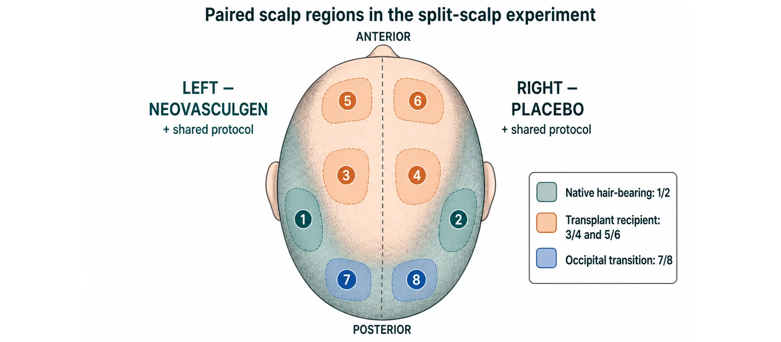
Four matched left-right regions were included. In every pair, the left region received Neovasculgen and the corresponding right region received placebo.
| Paired sites | Scalp region | Starting biological context |
|---|---|---|
| 1 and 2 | Lateral temporal-parietal region, approximately 4-5 cm above the ear | Native hair-bearing scalp without implanted grafts |
| 3 and 4 | Central parietal/vertex region | Recipient region of the second hair transplantation |
| 5 and 6 | Frontal region | Recipient region of the first hair transplantation |
| 7 and 8 | Occipital transition between visibly bald and hair-bearing scalp | Existing native follicles without implanted grafts; near the occipital donor field |
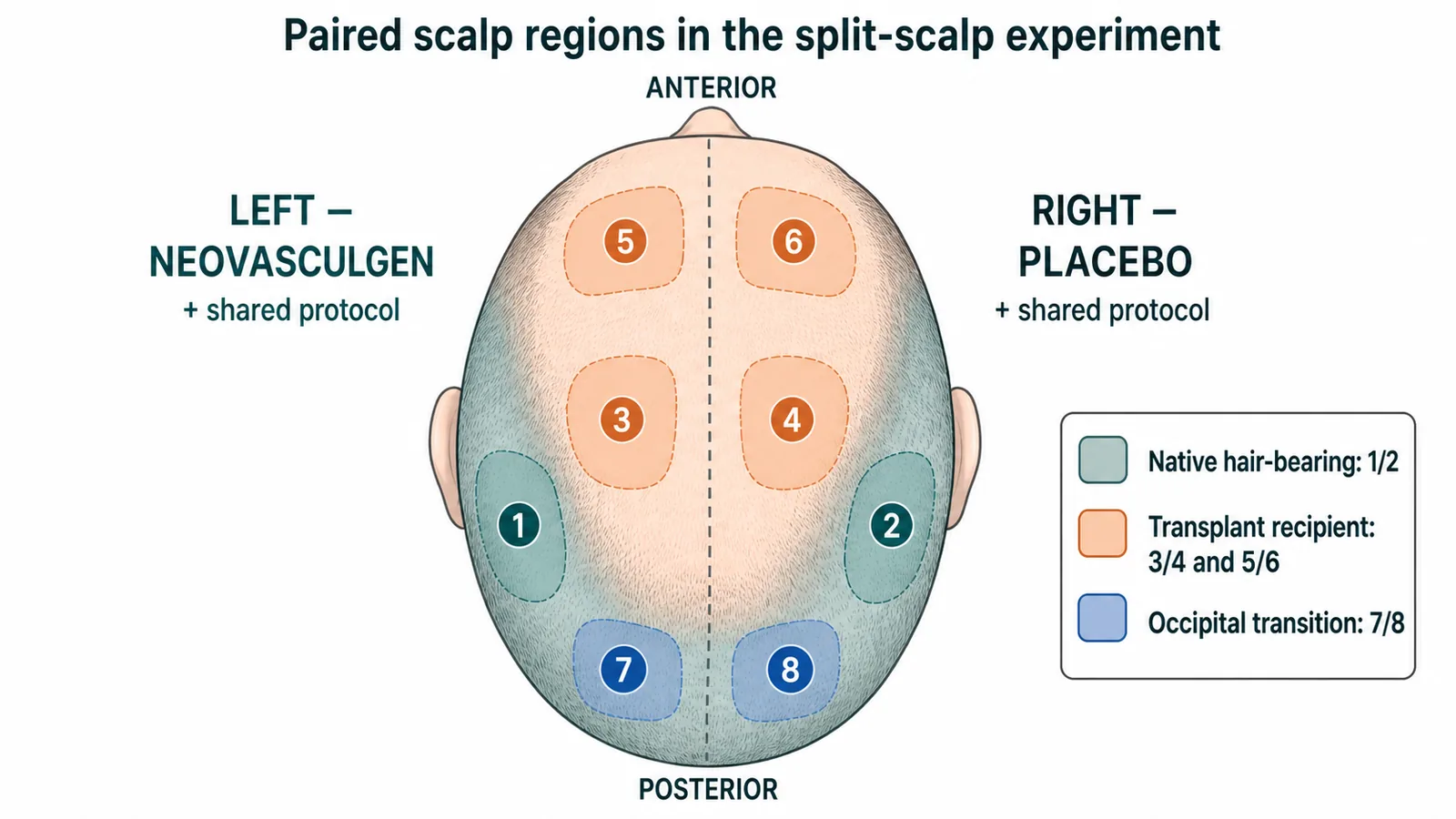
Figure 1. Site map of the blinded split-scalp experiment. Left scalp sites received VEGF165 plasmid therapy in addition to the shared background protocol; matched right-sided sites received placebo plus the same background protocol. The two non-transplanted pairs (1/2 and 7/8) were considered the most interpretable for the primary exploratory analysis.
Why We Focused on Two Regions
The four site pairs were not equally suitable for interpretation. Sites 3/4 and 5/6 were located in transplant recipient areas and included transplanted follicles. Measurements in these areas may have been affected by graft distribution, postoperative shedding, variable timing of hair emergence, and gradual maturation of transplanted shafts. These factors can influence hair density, anagen proportion, and shaft diameter independently of study treatment.
Results from the two transplant recipient pairs were mixed. No consistent between-side difference was observed for total density, mean diameter, and terminal-hair measurements. The current data cannot distinguish a treatment-related effect from expected changes during graft recovery and maturation. A repeat assessment of the transplant recipient areas should be performed approximately six months after the current visit, when postoperative changes are likely to be less prominent and graft growth and recipient-site healing are more stable.
For this reason, the clearest assessment of existing follicles came from the two pairs without implanted grafts:
- Sites 1/2: an already hair-bearing temporal-parietal region;
- Sites 7/8: the occipital transition between visibly bald and hair-bearing scalp.
These regions addressed the central biological question more directly: could local VEGF expression help follicles that were still present, but possibly thinning, miniaturized or functionally impaired, produce more visible or thicker hairs?
The Main Six-Month Result
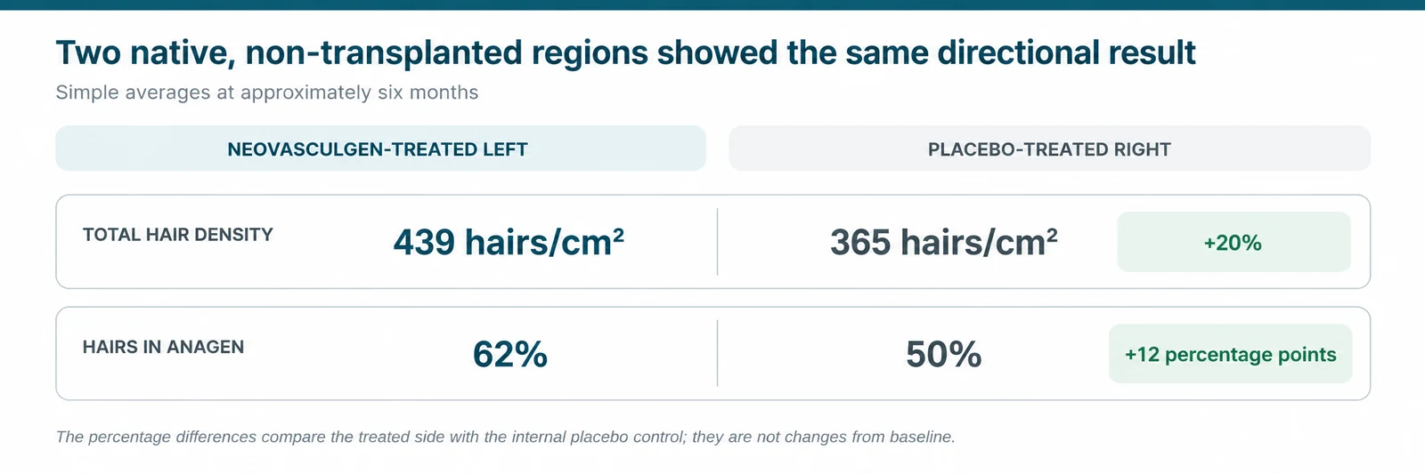
Figure 2. Simple endpoint averages across the two scalp regions without implanted grafts at approximately six months. The treated side showed 20% higher average total density and 12 percentage points more hairs in anagen than the internal placebo control. These values are descriptive endpoint comparisons; longitudinal changes for each region are presented separately below.
At approximately six months, both matched scalp pairs without implanted grafts showed the same two endpoint findings: the Neovasculgen-treated side had a higher total hair density and a higher proportion of hairs in anagen than the contralateral placebo side.
Across the two pairs, simple endpoint averages were 439 versus 365 hairs/cm², corresponding to 20% higher density on the treated side, and 62% versus 50% anagen, a difference of 12 percentage points.
In the temporal-parietal pair, density increased by 132 hairs/cm² on the treated side and by 58 hairs/cm² on the placebo side, producing a +74 hairs/cm² difference in change. Anagen increased by 12 percentage points on the treated side but decreased by 9 percentage points on the placebo side, yielding a +21-percentage-point difference in change.
In the occipital pair, anagen increased on both sides, with a more modest +5-percentage-point difference in change in favor of the treated site. The clearest longitudinal finding in this region was hair-shaft caliber: mean diameter increased by 4 μm at the treated site but decreased by 3 μm at the placebo site, producing a +7 μm difference in change.
The pattern of change therefore differed between the two regions. In the temporal-parietal pair, the larger between-side differences were observed for hair density and anagen proportion. Most of the additional hairs at the treated site were fine. In the occipital pair, the largest between-side difference was observed for mean hair-shaft diameter.
These findings may reflect different responses of existing follicles in the two scalp regions. However, this was a single-participant study, and follow-up is needed to determine whether the observed differences persist.
Occipital pair, sites 7 and 8: a caliber signal
The occipital measurement pair was located at the transition between visibly bald and hair-bearing scalp. This boundary may be particularly informative because it is likely to contain a heterogeneous population of follicles, including normal terminal follicles, miniaturized follicles producing fine hairs, and structurally preserved but functionally impaired follicles. Unlike long-standing completely bald scalp, such a transition zone may retain follicles capable of increasing their productive output in response to a supportive local intervention.
Here, the most informative result was the longitudinal change in mean hair diameter. Diameter increased from 33 to 37 μm at site 7 but decreased from 33 to 30 μm at site 8, producing a +7 μm between-side difference in change.
Table 1. Longitudinal changes in the occipital transition zone
| Measurement | Treated left | Placebo right | Between-side difference in change |
|---|---|---|---|
| Average diameter of all hairs | 33→37 μm; +4 μm (+12.1%) | 33→30 μm; −3 μm (−9.1%) | +7 μm |
| Total hair density | 179→570 hairs/cm²; +391 | 153→506 hairs/cm²; +353 | +38 hairs/cm² |
| Hairs in anagen | 21→48%; +27 percentage points | 18→40%; +22 percentage points | +5 percentage points |
| Terminal-hair density* | 90→327 hairs/cm²; +237 | 82→208 hairs/cm²; +126 | +111 hairs/cm²* |
| Terminal-hair proportion* | 50→58%; +8 percentage points | 53→41%; −12 percentage points | +20 percentage points* |
| Average diameter of terminal hairs* | 46→50 μm; +4 μm | 44→44 μm; 0 μm | +4 μm* |
Values are descriptive. The between-side difference in change was calculated as the change at the treated site minus the corresponding change at the placebo site. Positive values indicate a numerical difference in favor of the treated site.
* Terminal-hair outcomes should be considered exploratory because the classification threshold was not standardized across assessments. Terminal hairs were defined as >30 μm in November 2025, whereas the August 2026 reports used >35 μm at site 01 and >30 μm at site 02.
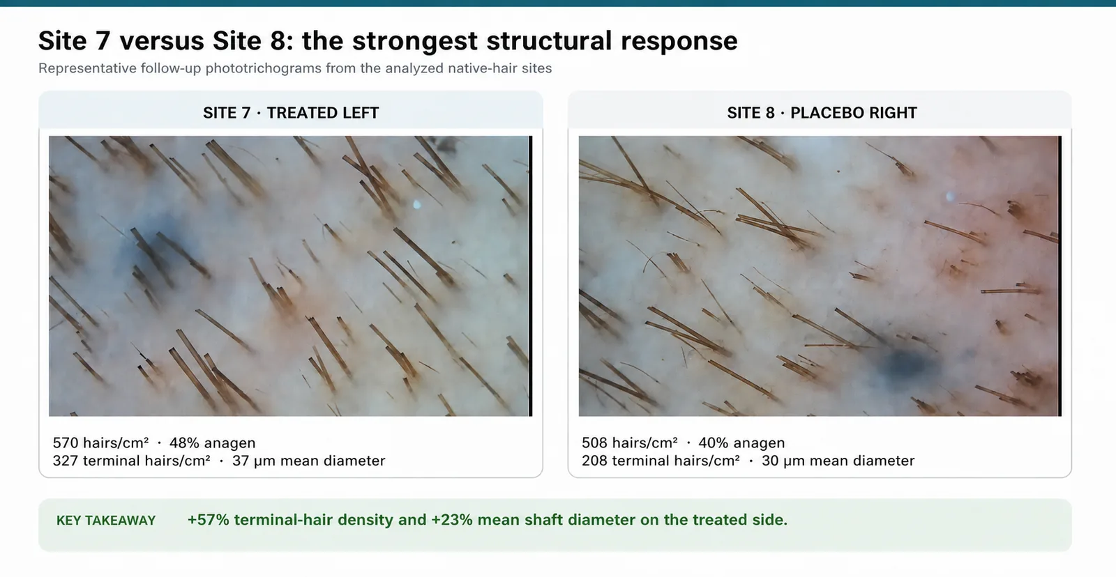
Figure 3. Follow-up phototrichograms of site 7 (Neovasculgen-treated left) and site 8 (placebo-treated right), acquired August 13, 2026. Quantitative conclusions are based on software-derived measurements; the images provide visual context.
Total hair density increased markedly on both sides. Although the absolute increase was 38 hairs/cm² greater at the treated site, the relative increase was not greater: density increased by 218.4% on the treated side and by 230.7% on the placebo side. Total density therefore does not provide a clear treatment-specific signal in this pair.
The proportion of hairs in anagen also increased on both sides, by 27 percentage points at the treated site and by 22 percentage points at the placebo site. The resulting between-side difference of +5 percentage points was modest and should be regarded as supportive rather than definitive.
Terminal-hair density increased by 237 hairs/cm² at the treated site and by 126 hairs/cm² at the placebo site, a between-side difference of 111 hairs/cm². This difference does not represent 111 newly formed follicles. Terminal-hair density is determined both by the number of visible hairs and by the number of hairs that meet the diameter criterion for terminal hair. An increase in this measure may therefore reflect thickening of existing fine hairs. Interpretation is limited because the diameter threshold used to classify terminal hairs differed between assessments.
Because progressive reduction in shaft caliber is central to follicular miniaturization in androgenetic alopecia, this finding is consistent with improved hair production by surviving follicles at the treated site. It does not establish follicular neogenesis or definitive reversal of miniaturization.
Why diameter matters
Follicular miniaturization is a key pathological feature of androgenetic alopecia. Affected follicles produce progressively thinner and shorter hair shafts, and anagen duration may decrease over successive hair cycles. Visible thinning can therefore reflect both a reduction in the number of growing hairs and a reduction in the diameter of hairs produced by surviving follicles [8].
Hair-diameter variability is one of the most established trichoscopic markers of this process. De Lacharrière and colleagues found that variation in hair diameter was the clinical parameter most closely associated with histological follicular miniaturization. A 2024 systematic review similarly reported hair-diameter variability in approximately 94% of patients with androgenetic alopecia, identifying it as the most frequently observed trichoscopic feature of the condition [8,9].
Hair caliber may also change before conventional density measurements show a clear decline. Recent trichoscopic evidence suggests that male androgenetic alopecia is commonly characterized first by progressive reduction in hair diameter and subsequently by a reduction in the number of hairs per follicular unit. Diameter may therefore be particularly useful when examining a transition zone in which follicles remain present but have begun to produce finer shafts [10].
The occipital transition zone likely contained existing follicles producing fine hairs. The increase in mean shaft diameter at the treated site may reflect a change in the output of these follicles, although the mechanism cannot be determined from this study.
In mice, increased VEGF expression has been associated with greater perifollicular vascularization, faster hair regrowth, and larger follicles and hair shafts. These preclinical findings provide a rationale for studying VEGF in human hair loss, but cannot be used to infer clinical efficacy [1].
Temporal-parietal pair, sites 1 and 2: more hairs entering active growth
The temporal-parietal pair represented native hair-bearing scalp without implanted grafts.
From November 2025 to August 2026, total hair density increased more than twice as much at the Neovasculgen-treated temporal-parietal site as at the placebo site.
Quantitative baseline measurements showed that the two sites had similar total hair density in November 2025: 175 hairs/cm² at the Neovasculgen-treated left site and 165 hairs/cm² at the placebo-treated right site. By August 2026, density had increased to 307 and 223 hairs/cm², respectively. This corresponded to an increase of 132 hairs/cm² on the treated side and 58 hairs/cm² on the placebo side, a between-side difference in change of +74 hairs/cm².
The anagen findings showed an even clearer directional shift. At baseline, the treated site had a slightly lower anagen proportion than the placebo site, at 64% versus 69%. At follow-up, the relationship was reversed: 76% of hairs were in anagen at the treated site compared with 60% at the placebo site. Anagen therefore increased by 12 percentage points on the treated side but decreased by 9 percentage points on the placebo side, producing a between-side difference in change of +21 percentage points.
Table 2. Longitudinal changes in the temporal-parietal pair
| Measurement | Site 1: treated left, November→August | Site 2: placebo right, November→August | Between-side difference in change |
|---|---|---|---|
| Total hair density | 175→307 hairs/cm²; +132 | 165→223 hairs/cm²; +58 | +74 hairs/cm² |
| Hairs in anagen | 64→76%; +12 percentage points | 69→60%; −9 percentage points | +21 percentage points |
| Average diameter of all hairs | 39→39 μm; no change | 48→46 μm; −2 μm | +2 μm |
| Terminal-hair density* | 120→144 hairs/cm²; +24 | 140→154 hairs/cm²; +14 | +10 hairs/cm²* |
| Thin-hair density* | 55→164 hairs/cm²; +109 | 25→69 hairs/cm²; +44 | +65 hairs/cm²* |
| Terminal-hair proportion* | 69→47%; −22 percentage points | 85→69%; −16 percentage points | −6 percentage points* |
| Average terminal-hair diameter* | 47→61 μm; +14 μm | 53→59 μm; +6 μm | +8 μm* |
The between-side difference in change was calculated as the change at the treated site minus the corresponding change at the placebo site. Positive values favor the treated site.
* Terminal- and thin-hair outcomes are exploratory because the classification threshold was not fully standardized. At the treated baseline site, terminal hairs were defined as >30 μm, whereas a >35 μm threshold was used at the placebo baseline site and at both follow-up sites. Total density, anagen proportion, and mean diameter of all hairs are not affected by this classification difference.
The increase in total density was composed predominantly of hairs classified as fine at follow-up. However, the exact change in fine- and terminal-hair density cannot be interpreted as biological conversion because the classification threshold changed at the treated site. More importantly, the threshold-independent measure of total hair density increased considerably more on the treated side.
Mean diameter across all hairs remained stable at 39 μm at the treated site, despite the marked increase in the number of visible hairs. At the placebo site, mean diameter decreased slightly from 48 to 46 μm. The treated site therefore did not show an increase in overall shaft caliber, but it maintained its baseline mean diameter while adding a substantial population of visible, predominantly fine hairs.
Taken together, the temporal-parietal findings are consistent with increased visible hair production and greater anagen activity at the Neovasculgen-treated site. One possible interpretation is that more existing follicles entered productive growth, while many of the additional hairs had not yet reached a mature terminal caliber. The data do not establish the formation of new follicles or prove reactivation of dormant follicles. Continued follow-up will be needed to determine whether the additional fine hairs persist and develop into thicker shafts.
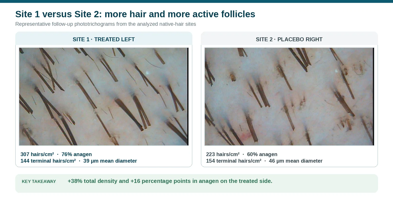
Figure 4. Follow-up phototrichograms of the temporal-parietal pair: site 1, Neovasculgen-treated left (August 2026), and site 2, placebo-treated right (August 2026). From November 2025 to August 2026, total density increased by 132 hairs/cm² at site 1 and by 58 hairs/cm² at site 2. Anagen increased by 12 percentage points at site 1 but decreased by 9 percentage points at site 2. The additional hair population at the treated site consisted predominantly of fine hairs.
What Happened in the Transplanted Areas?
Transplant recipient areas were also assessed, but the results were more difficult to interpret than those from the non-transplanted sites. In the region assessed approximately nine months after transplantation, total visible hair density was higher on the treated side, whereas terminal-hair density and mean hair diameter were higher on the placebo side. In the region assessed approximately three months after transplantation, the placebo side had a greater proportion of mature terminal hairs at that time point.
Measurements obtained during the first months after transplantation are influenced by postoperative shedding, variable timing of graft re-entry into anagen, gradual emergence of transplanted hairs, and subsequent shaft maturation. These processes can affect density, anagen proportion, and hair diameter independently of the study treatment. The transplant recipient sites therefore cannot yet be used to assess a treatment-related effect of Neovasculgen.
Repeat phototrichogram measurements should be obtained approximately six months after the current assessment. By that time, early postoperative changes should have largely passed, more grafts should have entered stable visible growth, and the recipient tissue should be more fully healed. A later assessment will provide a more appropriate basis for comparing the treated and placebo sides in the transplant areas.
How Does This Compare With Existing Hair-Loss Treatments?
Direct comparisons between this N-of-1 experiment and published trials should be made cautiously. Hair studies differ in treatment duration, scalp region, disease severity, background therapy, measurement method, and definition of terminal hair. Nevertheless, published results for minoxidil, platelet-rich plasma (PRP), and mesotherapy provide useful context for the magnitude and type of changes that we observed.
| Treatment | Hair density | Hair-shaft diameter | Anagen | Strength of evidence |
|---|---|---|---|---|
| Topical minoxidil | In a 48-week randomized trial, 5% minoxidil increased non-vellus hair count by 18.6 hairs/cm² from baseline, compared with 3.9 hairs/cm² with vehicle | Diameter can increase, but it is not consistently reported in pivotal trials; a modern comparative study reported an increase of approximately 3 μm with topical minoxidil over six months | One six-month study reported an 11.68% improvement in the anagen ratio with 5% minoxidil | Established treatment supported by multiple randomized trials |
| PRP | Meta-analyses suggest an average advantage of approximately 25-28 hairs/cm² over control, although results are highly heterogeneous | Meta-analyses have not demonstrated a consistent significant benefit; one analysis estimated a mean difference of 2.02 μm, with a confidence interval crossing zero | Several split-scalp studies reported increased anagen hair, but the size of the effect is not reported consistently | Moderate but heterogeneous evidence |
| Present split-scalp experiment: Neovasculgen plus shared protocol versus placebo plus shared protocol | Endpoint density favored the treated side in both pairs. Sites 1/2: 307 vs 223 hairs/cm²; sites 7/8: 570 vs 506 hairs/cm². The simple endpoint average was 439 vs 365 hairs/cm² (+20.3%). Longitudinally, density increased by 132 vs 58 hairs/cm² at sites 1 vs 2 (+74 hairs/cm² difference in change). At sites 7 vs 8, density increased by 391 vs 353 hairs/cm² (+38 hairs/cm² difference in change) | The pattern was region-dependent. At sites 1/2, mean diameter remained stable at 39 μm on the treated side and decreased from 48 to 46 μm on the placebo side (+2 μm difference in change). At sites 7/8, diameter increased from 33 to 37 μm on the treated side and decreased from 33 to 30 μm on the placebo side (+7 μm difference in change) | Anagen showed more favorable longitudinal change on the treated side in both pairs. Sites 1 vs 2: 64→76% vs 69→60% (+21-percentage-point difference in change). Sites 7 vs 8: 21→48% vs 18→40% (+5-percentage-point difference in change). The endpoint average was 62% vs 50% | Exploratory blinded N-of-1 split-scalp observation |
Minoxidil
Minoxidil has the strongest evidence base of the three comparators. In a 48-week randomized trial involving 393 men, 5% topical minoxidil increased non-vellus hair count by 18.6 hairs/cm² from baseline, compared with 12.7 hairs/cm² with 2% minoxidil and 3.9 hairs/cm² with vehicle. The 5% formulation produced an earlier and greater response than the 2% formulation [11].
Minoxidil was used on both sides of the scalp throughout the study, together with topical finasteride and the other background treatments. The side-to-side comparison therefore assessed Neovasculgen plus background treatment versus placebo plus the same background treatment. It did not compare Neovasculgen with minoxidil or finasteride alone.
Platelet-rich plasma
PRP is the most relevant established injectable comparator. A meta-analysis of 10 randomized trials found approximately 25 additional hairs/cm² with PRP versus control, but no statistically significant overall effect on hair diameter. Another meta-analysis estimated a density difference of 27.55 hairs/cm², although heterogeneity was high and the certainty of evidence was rated low; its estimated diameter difference of 2.02 μm was not conclusive [12].
These findings suggest that PRP has its most consistent effect on density, while changes in shaft caliber are less reproducible. In the present experiment, the temporal-parietal pair also showed a density-dominant pattern, whereas the occipital pair showed a clearer diameter signal. This comparison is descriptive only and does not establish equal or superior efficacy.
Combination treatment
A meta-analysis of six studies reported that PRP combined with minoxidil produced approximately 9.14 additional hairs/cm² and a 4.72 μm greater increase in diameter than minoxidil or PRP alone. This is relevant because regenerative interventions may be most realistically developed as add-on treatments rather than replacements for established therapy [13].
The current split-scalp experiment followed the same add-on logic: both sides received minoxidil, finasteride, red-light therapy and matched injections, while Neovasculgen was the intended local difference.
Across the two evaluated non-recipient regions, the Neovasculgen-treated side on top of other therapies showed a 20% higher simple average density and 12 percentage points more anagen at follow-up. These differences appear numerically larger than the average density advantages reported in many minoxidil and PRP trials, but the values are not directly comparable: the present results are cross-sectional side-to-side differences from one participant, not group-level treatment effects or standardized changes from baseline.
The more important observation is the consistency of direction across two anatomical regions and the complementary nature of the response: more fine growing hairs in the temporal-parietal region and greater shaft caliber in the occipital transition zone. This pattern is biologically interesting, but confirmation will require a prospective study with standardized field relocation, uniform hair-classification thresholds and multiple participants.
What This Suggests for Future Patient Studies
1. Patients who do not plan to undergo transplantation
This is the most directly supported development group. The clearest positive signal appeared in native hair-bearing regions not affected by transplantation. The intended target would be follicles that are still present but thinning or miniaturized, not long-bald skin without functional follicles.
2. Transplant patients who want to preserve their remaining native hair
Hair transplantation redistributes follicles but does not stop continuing miniaturization of surrounding native hair. Neovasculgen could be evaluated in the native-hair areas around the transplant field as an addition to minoxidil, finasteride, and other preservation measures.
3. Direct treatment of the recipient zone
Administration into the recipient area, including after transplantation, remains biologically plausible because grafts must integrate into new tissue and establish vascular support. The current experiment does not isolate an effect on transplantation outcomes, so this question requires its own controlled study.
4. Patients concerned about progressive hair thinning and hair quality
Future studies could also include patients who have early signs of follicular miniaturization, such as reduced hair-shaft diameter, increased hair-diameter variability, or a gradual decline in scalp coverage. The aim would be to evaluate whether treatment can help preserve the output of existing follicles before more advanced hair loss develops.
Conclusion
Minoxidil and finasteride are commonly used treatments for androgenetic alopecia, but some patients continue to lose hair or achieve only a partial response. Additional treatments are needed, particularly for patients with ongoing thinning despite standard therapy.
In this blinded split-scalp N-of-1 study, VEGF165 plasmid therapy (Neovasculgen) was administered to one side of the scalp. The contralateral side received placebo. Both sides continued topical minoxidil, topical finasteride, daily red-light therapy, scalp massage, and the same injection procedure.
In both non-transplanted site pairs, total hair density and anagen proportion were higher at the Neovasculgen-treated site at follow-up than at the matched placebo site. In the temporal-parietal pair, the treated site showed a larger increase in hair density and anagen proportion; most of the additional hairs were fine. In the occipital pair, mean hair-shaft diameter increased at the treated site and decreased at the placebo site, resulting in a 7 μm between-side difference in change.
These observations do not prove that VEGF therapy generated new follicles, reversed miniaturization or outperformed established treatments. They do, however, support further investigation of local VEGF plasmid therapy as a possible add-on approach for scalp regions where follicles remain present but are thinning or functionally impaired.
VEGF gene therapy is experimental and is not approved by the FDA for hair loss. Unlimited Bio is not a medical provider; treatment is administered by independent clinics. This article describes a single-participant observation and is not medical advice.
Selected References
- Yano K, Brown LF, Detmar M. Control of hair growth and follicle size by VEGF-mediated angiogenesis. Journal of Clinical Investigation. 2001;107(4):409-417. doi:10.1172/JCI11317
- Gupta AK, Venkataraman M, Talukder M, Bamimore MA. Relative efficacy of minoxidil and the 5-alpha reductase inhibitors in androgenetic alopecia treatment of male patients: a network meta-analysis. JAMA Dermatology. 2022;158(3):266-274. doi:10.1001/jamadermatol.2021.5743
- Rossi A, Caro G. Efficacy of the association of topical minoxidil and topical finasteride compared with their use in monotherapy in men with androgenetic alopecia. Journal of Cosmetic Dermatology. 2024. doi:10.1111/jocd.15953
- Suchonwanit P, Srisuwanwattana P, Chalermroj N, Khunkhet S. Topical finasteride combined with minoxidil versus minoxidil alone in male androgenetic alopecia. Journal of the European Academy of Dermatology and Venereology. 2018;32(12):2257-2263. doi:10.1111/jdv.15171
- Huang CH, Fu Y, Chi CC. Health-related quality of life, depression, and self-esteem in patients with androgenetic alopecia: a systematic review and meta-analysis. JAMA Dermatology. 2021.
- Deev RV, Bozo IY, Mzhavanadze ND, et al. pCMV-vegf165 intramuscular gene transfer is an effective method of treatment for patients with chronic lower limb ischemia. Journal of Cardiovascular Pharmacology and Therapeutics. 2015;20(5):473-482. doi:10.1177/1074248415574336
- Morgunov I. VEGF gene therapy for hair loss, tested on half of our CEO’s scalp. Unlimited Bio, February 23, 2026. unlimited.bio/blog
- Kuczara A, Waśkiel-Burnat A, Rakowska A, Olszewska M, Rudnicka L. Trichoscopy of androgenetic alopecia: a systematic review. Journal of Clinical Medicine. 2024;13(7):1962. doi:10.3390/jcm13071962
- de Lacharrière O, Deloche C, Misciali C, et al. Hair diameter diversity: a clinical sign reflecting the follicle miniaturization. Archives of Dermatology. 2001;137(5):641-646.
- Kamishima T, Hirabe C, Myint KZY, Taguchi J. Divergent progression pathways in male androgenetic alopecia and female pattern hair loss: trichoscopic perspectives. Journal of Cosmetic Dermatology. 2024;23:1828-1839. doi:10.1111/jocd.16177
- Olsen EA, Dunlap FE, Funicella T, et al. A randomized clinical trial of 5% topical minoxidil versus 2% topical minoxidil and placebo in the treatment of androgenetic alopecia in men. Journal of the American Academy of Dermatology. 2002;47(3):377-385. doi:10.1067/mjd.2002.124088
- Li M, Qu K, Lei Q, Chen M, Bian D. Effectiveness of platelet-rich plasma in the treatment of androgenic alopecia: a meta-analysis. Aesthetic Plastic Surgery. 2024;48(5):977-984. doi:10.1007/s00266-023-03603-9
- Xiao C, Zhang GH, Li HQ, Yang PP, Zhang HB, Mu YX. Meta-analysis of efficacy of platelet-rich plasma combined with minoxidil for androgenetic alopecia. Aesthetic Plastic Surgery. 2024;48(21):4554-4566. doi:10.1007/s00266-024-04054-6
Ivan Morgunov